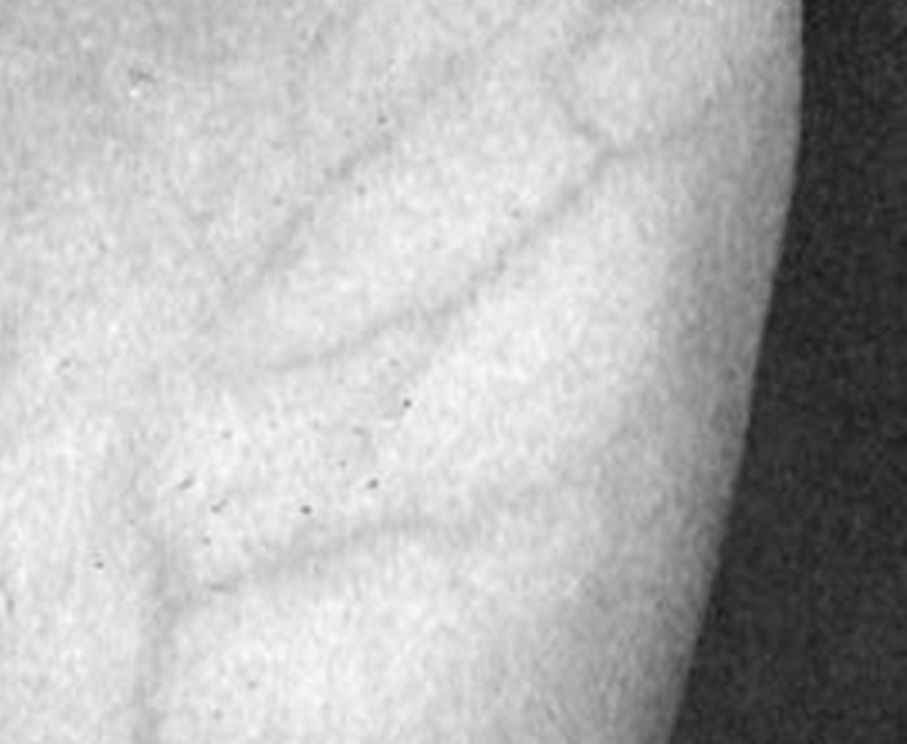
Background: Lipedema is characterized by the deposition of abnormal fat in the lower and upper limbs bilaterally. It is a disease with high prevalence and genetic characteristics. Non-specific and non-quantified increases in the thickness of the subcutaneous tissue have previously been demonstrated using magnetic resonance imaging and computed tomography.
Objectives: To evaluate the thickness of the dermis and subcutaneous tissue in predetermined areas as a distinguishing feature between individuals with and without lipedema using ultrasound.
Methods: Ultrasound images of 89 female patients were analyzed, including patients undergoing clinical investigation for venous insufficiency or lipedema who underwent ultrasound evaluations at our institution. Patients were divided in two groups: with lipedema clinically diagnosed and those without lipedema. They underwent a common Doppler protocol for venous mapping to assess venous insufficiency associated with the evaluation of dermis and subcutaneous thickness at pre-defined points of the lower limbs.
Results: There were 63 patients with lipedema. Anterior thigh, pre-tibial and lateral aspect of the leg and supra-just medial malleolar region were significantly different. Supra-just medial malleolar region was significantly different with BMI above 25. An optimal cutoff value was calculated for the ultrasound diagnosis of lipedema using thickness of the dermis and subcutaneous tissues.
Conclusions: Studied criteria allow use of simple and reproducible ultrasound cutoff values to diagnose lipedema in the lower limbs. Pre-tibial region thickness measurement, followed by thigh and lateral leg thickness are recommended for the ultrasound diagnosis of lipedema.

Você também pode se interessar por:
Livros publicados pelo Dr. Alexandre Amato
Para todos: Dieta Anti-inflamatória Estratégica: a sua dieta pessoal. Complete seu conhecimento nutricional com este livro. Cansada(o) de procurar a dieta ideal? Já pensou… Continue a ler »Livros publicados pelo Dr. Alexandre Amato
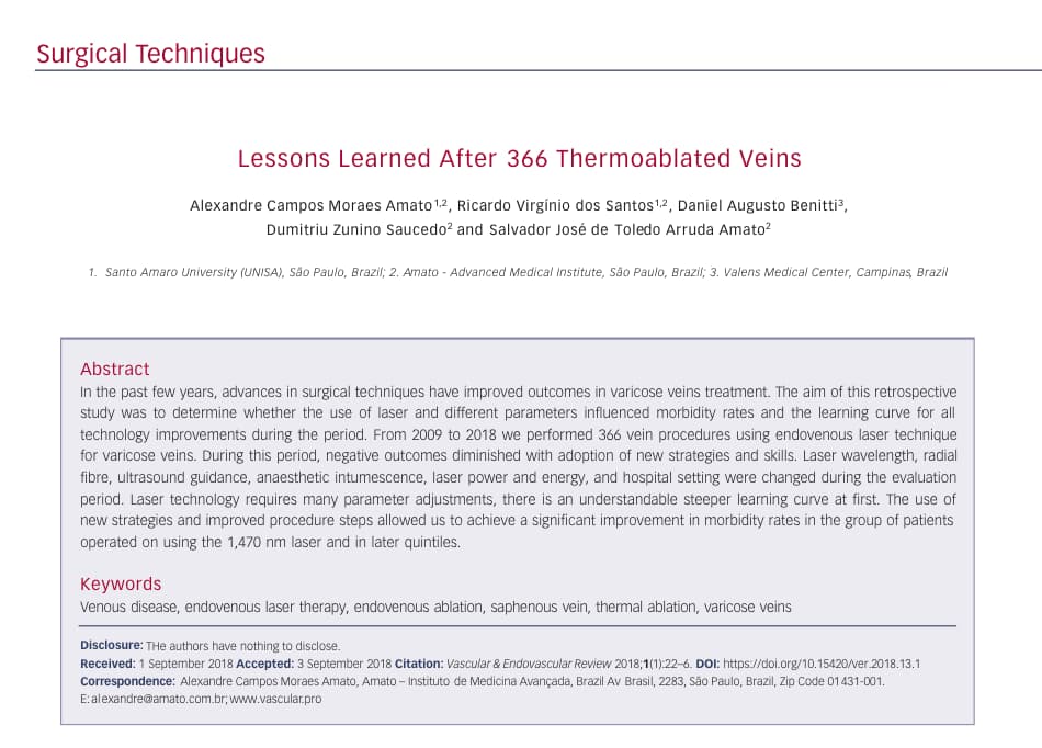
Lessons learned after 366 thermoablated veins
A equipe vascular.pro publicou artico sobre cirurgia de varizes com laser em revista internacional. Abstract: In the last few years, advances in surgical techniques have… Continue a ler »Lessons learned after 366 thermoablated veins
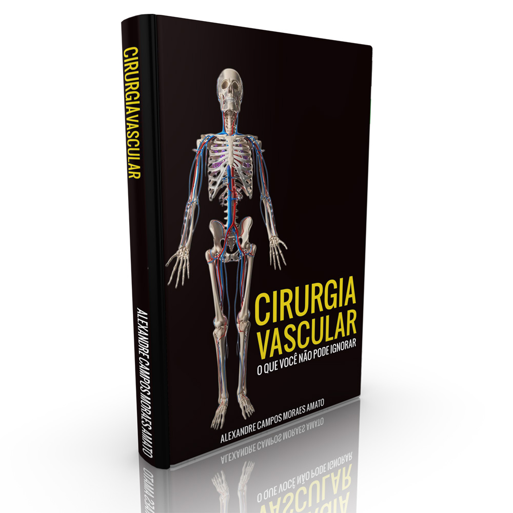
Livro: Vascular. O que você não pode ignorar.
Livro recomendado para a disciplina de Cirurgia Vascular da UNISA.
VeinCamera: Foto identificador de veias varicosas
O Dr Alexandre Amato criou aplicativo para iPad (também roda no iPhone) para visualização de veias (varicosas ou não) pela câmera do iPad. O aplicativo… Continue a ler »VeinCamera: Foto identificador de veias varicosas
Predictors of Adamkiewicz artery and anterior spinal artery detection through computerized tomographic angiography
Abstract Background: The detection of the Adamkiewicz artery and the anterior spinal artery has been associated with the ability to prevent adverse spinal cord outcomes… Continue a ler »Predictors of Adamkiewicz artery and anterior spinal artery detection through computerized tomographic angiography